
Multiple sclerosis (MS) is characterized by central nervous system white matter lesions that affect people’s ability to move independently. Further, people with MS often report significant asymmetries in muscle strength and function in the left versus the right leg. These are often associated with increased postural sway (i.e. worse balance) and less symmetrical stepping patterns during walking (i.e. worse walking). It is no surprise that such lower limb asymmetries are frequently associated with poorer balance control, falls, and reduced quality of life. Currently there is limited understanding as to why these limb asymmetries exist in MS and which areas within the central nervous system contribute to these mobility-limiting issues. It is also unknown whether improving these lower limb asymmetries may concomitantly improve balance and mobility during activities of daily living.
To address these questions, participants stood on a platform and tried to maintain their balance while the platform continually slid forward and backward at a fixed frequency of differing amplitudes. We measured their ability to anticipate changes in direction (i.e. temporal performance) and their ability to control the amplitude of sway (i.e. spatial performance) with repeated exposures to this moving platform. To understand the neural underpinnings of postural motor learning, we correlated the acquisition and retention of practice-related improvements in postural control to brain white matter microstructural integrity acquired via diffusion weighted magnetic resonance images using a tract-based spatial statistical approach. Despite having worse postural control than control participants, those with MS exhibited improvements in temporal performance (over one day of practice) and retention (ability to maintain improvements 24 hours later) in a similar manner as control participants. Improvements in temporal performance were directly correlated to microstructural integrity of white matter tracts in the corpus callosum, posterior parieto-sensorimotor fibers and the brainstem in people with MS. Within the corpus callosum, fibers connecting the primary motor cortices (red fibers in Figure 1)were most strongly correlated to temporal improvements in postural control, in contrast to those connecting pre-supplementary or supplementary motor areas (yellow and orange fibers in Figure 1).
For movements that require precise coordination between the two sides of the body (e.g. walking, postural control of balance, typing) a delicate balance of excitation and inhibition is required between the right and left sensorimotor cortices. This interhemispheric communication is principally accomplished through the corpus callosum. Reduced quality of the corpus callosum is common in people with MS and has been directly related to poorer communication between the two sides of the brain and upper extremity motor performance. We suggest that impairments in gait and balance control are also, at least in part, a result of reduced structure and altered communication between the two sides of the brain in people with MS. However, our understanding of how changes in communication between the two sides of the brain contribute to lower limb asymmetries and the resultant declines in mobility for those with MS remains incomplete.
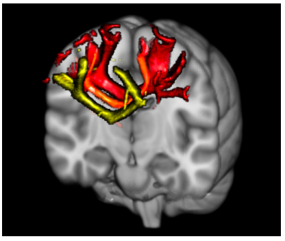
Figure 1 – Interhemispheric white matter fiber tracts connecting the right and left pre-supplementary motor areas (yellow), supplementary motor areas (orange), and primary motor cortices (red).
Copyright:
The ISPGR blog applied Creative Commons Attribution-ShareAlike 4.0 International (CC BY-SA 4.0) license to figure and text of the article.
https://creativecommons.org/licenses/by-sa/4.0/
Publication:
Daniel S. Peterson, Geetanjali Gera, Fay B. Horak, Brett W. Fling. Corpus Callosum Structural Integrity Is Associated With Postural Control Improvement in Persons With Multiple Sclerosis Who Have Minimal Disability. Neurorehabilitation and Neural Repair, Vol 31, Issue 4, pp. 343 – 353
http://journals.sagepub.com/doi/abs/10.1177/1545968316680487
About the Author

Brett W. Fling, Ph.D.
Director – Sensorimotor Neuroimaging Laboratory. Colorado State University
Brett W. Fling, Ph.D. Assistant Professor – Health and Exercise Science Department & Molecular, Cellular & Integrative Neurosciences Program. Director – Sensorimotor Neuroimaging Laboratory. Colorado State University, Fort Collins, Colorado.
Research within the Sensorimotor Neuroimaging Laboratory at Colorado State University is designed to understand the contributions of the brain’s structural and functional neural networks to everyday movements. We leverage this understanding of the nervous system to develop new therapeutic interventions for individuals with sensorimotor dysfunction. Our laboratory utilizes a range of neuroimaging techniques including functional and structural magnetic resonance imaging, diffusion tensor imaging, electroencephalography, and transcranial magnetic stimulation to assess neuroanatomy and neurophysiologic function. These state of the art imaging techniques are integrated with experimental paradigms relying on the biomechanical analysis of sensorimotor control to provide a comprehensive view of the neural control of movement.
Copyright
© 2018 by the author. Except as otherwise noted, the ISPGR blog, including its text and figures, is licensed under a Creative Commons Attribution-ShareAlike 4.0 International License. To view a copy of this license, visit https://creativecommons.org/licenses/by-sa/4.0/legalcode.
ISPGR blog (ISSN 2561-4703)
Are you interested in writing a blog post for the ISPGR website? If so, please email the ISGPR Secretariat with the following information:
- First and Last Name
- Institution/Affiliation
- Paper you will be referencing